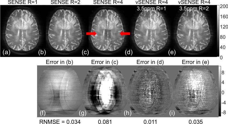
Figure 1.

Comparison of source images at 14ppm from a brain tumor patient reconstructed with conventional SENSE (R=1 in a, R=2 in b, and R=4 in c) and vSENSE using one 3.5ppm frame (fully-sample in d, and undersampled by a factor of 2 in e) to improve sensitivity maps. The difference of accelerated SENSE and vSENSE images (b–e) from the full k-space image (a) were shown in (f–i), respectively. Red arrows indicated artifacts in the conventional SENSE R=4 image. RNMSE stands for root normalized mean squared error.
