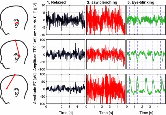
Fig. 3.

Time domain examples of recordings from a single subject. The first row shows recordings from the ELE-ELB electrode pair. The second and third row are recordings from the TP9-Cz and F7-Cz electrode pairs, respectively. The sketches on the left illustrate the electrode positions. The plots show raw EEG data band-pass filtered from 1 to 40 Hz. The blue dashed lines for the eye-blinking condition indicate the eye-blink cue
