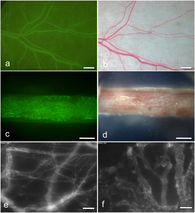
Fig 2. Intravital fluorescence microscopy (IVM) images.
(a) IVM of microvessels in the dorsal skinfold chamber (1.25× objective) and in the femur window (c) (2.5× objective) after contrast enhancement by 0.1 ml of 5% FITC-labeled dextran 150.000 i.v. (b;d) Stereo microscopy images of microvessels in the dorsal skinfold chamber (b) and in the femur window (d). (e;f) Intravital fluorescence microscopy of microvessels in the dorsal skinfold chamber (e) and microvessels in the femur window (f) (20× objective) by blue light epi-illumination. (scale bars a-b = 680 μm, c-d = 500 μm, e-f = 70 μm).

