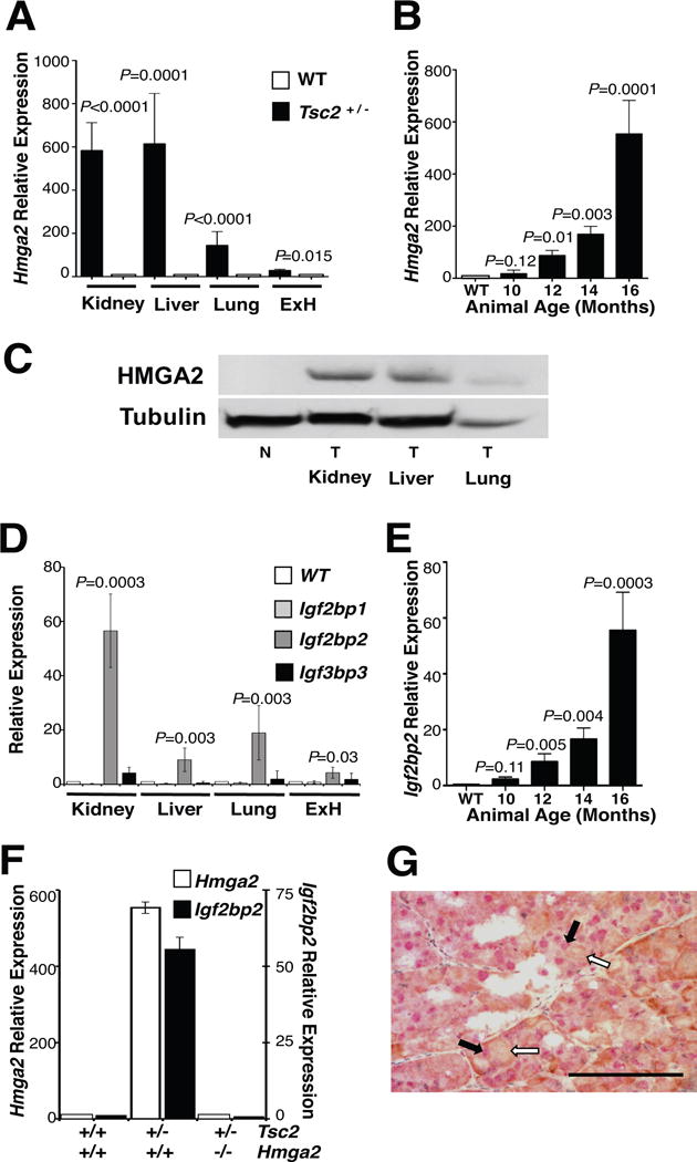
Figure 1. HMGA2 is expressed in all tumors from Tsc2+/− mice.

(A) Hmga2 is expressed in all tumors from 16 month-old Tsc2+/− mice. qPCR was performed on total RNA from tumors in multiple organs and corresponding normal tissue. (B) Hmga2 expression increases in kidney tumors as mice age. (C) Western blot performed on normal and tumor tissue from several organs for HMGA2 and tubulin expression. N, normal tissue; T, tumor. (D) Igf2bp2 was the only member of the Igf2bp family of genes to be expressed in 16-month-old Tsc2+/− mice tumors. (E) Igf2bp2 expression increased in renal tumors, as mice aged. (F) Igf2bp2 expression correlated with Hmga2 expression in renal tumors from 16-month-old mice for each genotype. (A-B, D-F) No difference was observed between Tsc2+/–Hmga2+/+ and Tsc2+/–Hmga2+/− mice. Error bars, mean + s.e.m. P value shown, comparing to normal tissue of corresponding organ. (G) Renal cell carcinoma double stained for nuclear HMGA2 (red) and cytoplasmic IGF2BP2 (brown). Black and white filled arrows denote positive nuclear HMGA2 staining (red staining) and positive cytoplasmic IGF2BP2 staining (brown staining), respectively. (Scale bar, 100 mm). See also Supplemental Figure S1 for additional immunohistochemistry results.
