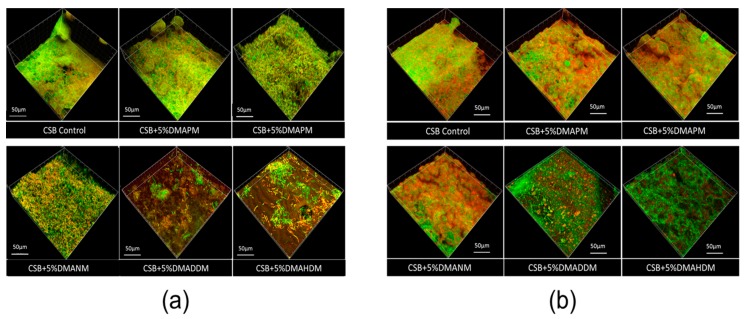
Figure 6.
Confocal laser scanning microscope (CLSM) of three-species biofilms. (a) Live/dead staining of biofilms on the adhesive disks of the six groups. Bacterial cells were stained green, and dead cells were stained red; (b) exopolysaccharide (EPS) staining of biofilms on the adhesive disks of the six groups. Bacteria were stained green, and EPS was stained red.

