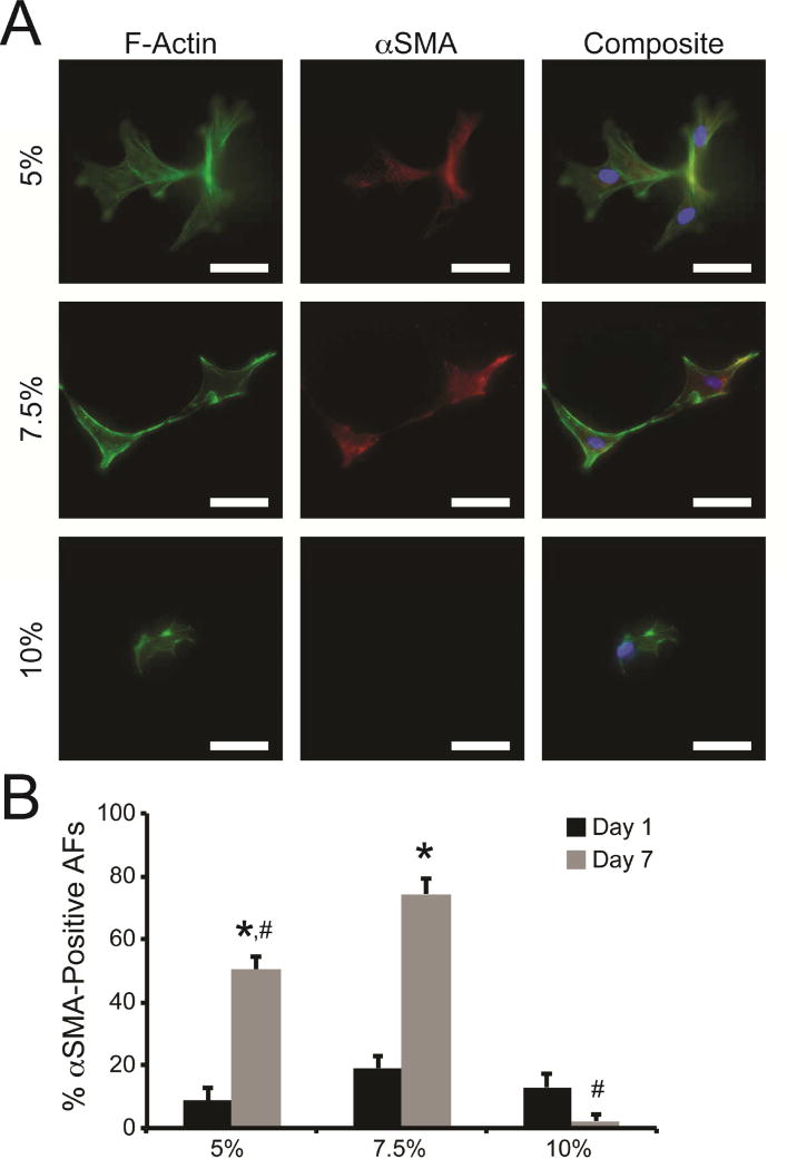
Fig. 4. Myofibroblast activation in response to hydrogel stiffness.
(A) Representative images of AFs cultured in 5%, 7.5%, and 10% hydrogels for 7 days. AFs were stained with antibodies for F-actin (green), αSMA (red), and nuclear stain Hoescht 33342 (blue). Scale bars = 50 µm. (B) Low levels of αSMA-positive AFs were detected in all hydrogels after 1 day of culture. After 7 days of culture, an increased number of AFs elicited αSMA staining in 5% and 7.5% hydrogels, while very few αSMA-positive cells were observed in 10% hydrogels. * represents significance from paired hydrogels on day 1; # represents significance from 7.5% hydrogels on day 7 (p<0.05 by two-way ANOVA with Tukey HSD).

