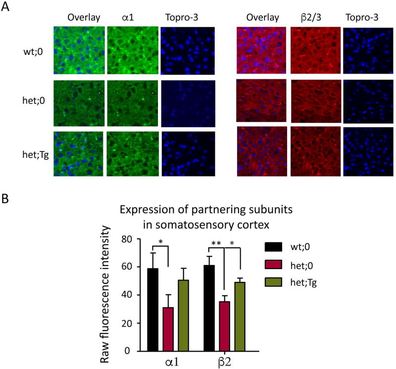
Figure 2. The expression levels of partnering α1 and β2/3 subunits were restored in the somatosensory cortex by the hGABRG2HA transgene.
A. Immunostaining of α1 and β2/3 subunits in somatosensory cortex of wt;0, het;0 and het;Tg mice was performed. Brains from 2 month old wt;0, het;0, and het;Tg mouse littermates were fixed and sectioned on a cryostat at 30 µm. The brain cortices were stained with anti-α1 or anti-β2/3 subunit antibody, and cortical layer VI was visualized. The nuclei were stained with TO-PRO-3 (blue). Images were acquired under confocal microscopy. B. Expression levels from 12 sections from 3 different mice for each genotype were plotted. The total raw fluorescence values of the α1 or β2/3 subunits in somatosensory cortex were quantified by ImageJ. (* p < 0.05; **p < 0.01 vs wt, *p< 0.05 vs het), unpaired t test).

