Figure 3.

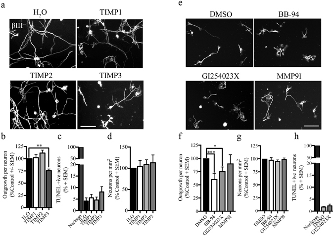
ADAM proteolytic activity is necessary for embryonic DRG growth. (a) Embryonic DRG neurons seeded on a PLL substrate and treated with control (H2O), or endogenous metalloproteinase inhibitors (TIMP1-3). Neuronal projections were visualized with βIII-tubulin staining. (b) Neurite outgrowth quantification of embryonic DRG neurons exposed to soluble recombinant TIMPs and control conditions. Data have been normalized to control condition. (c) TUNEL assay was performed in embryonic DRG neurons treated with TIMPs or H2O control. DNA nuclease was used as a positive control. Data are shown as a percentage of TUNEL-positive neurons. (d) Number of neurons present on the PLL substrate after treatment with soluble TIMPs and control condition. (e). Embryonic DRG neurons exposed to DMSO as a control, a selective ADAM10 inhibitor, GI254030X, BB-94 or a selective MMP9 inhibitor (MMP9I). (f–h) Quantification of neurite outgrowth (f), cell adhesion (g) and percentage TUNEL-positive neurons (h) following treatment with metalloproteinase inhibitors. For the TUNEL assay the data are shown as a percentage of TUNEL-positive neurons. For total number of neurons, data have been normalized to control DMSO or H2O. N = 3–4 from independent cultures. Data are shown as mean + S.E.M. *p < 0.05, **p < 0.01, ***P < 0.001by one-way ANOVA followed by Bonferroni post hoc test. Scale bar, 100 μm.
