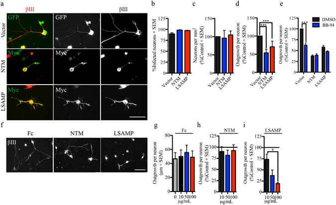
Figure 5.

Ectopic surface NTM and LSAMP repress neurite outgrowth in embryonic DRG neurons. (a) Embryonic (E18-19) DRG neurons seeded on a PLL substrate and expressing HSV-GFP or HSV-Myc-tagged IgLON constructs. Surface Myc-IgLON expression was detected with anti-Myc antibody in the absence of permeabilization. (b–e) Quantification of the number of infected neurons (b), total number of seeded neurons (c) and neurite outgrowth (d,e) from E18-19 DRG neurons expressing GFP (vector) or Myc-IgLON proteins in the presence or absence of BB-94. Data were normalized to control HSV-GFP. N = 3–6 from independent cultures. (f). βIII tubulin stained E18-19 DRG neurons plated on immobilized Fc or FC-IgLON substrates. (g–i) Quantification of outgrowth from embryonic DRG neurons seeded on immobilized Fc-IgLON substrates (10, 50 and 100 μg/mL). Data were normalized to control Fc substrate. N = 4 from independent cultures. Data are shown as mean + S.E.M. *p < 0.05, **p < 0.01, ***P < 0.001 by one-way ANOVA followed by Bonferroni post hoc test. Scale bar, 50 μm.
