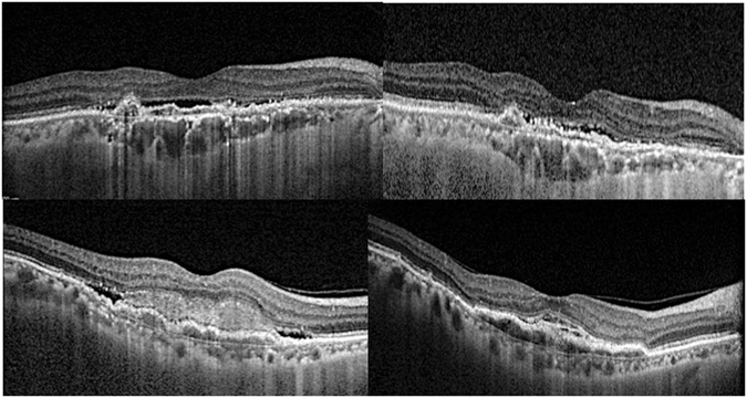
Figure 2.

The choroidal vascularity index (CVI), total choroidal area (TCA), luminal area (LA) and stromal area (SA) changes of neovascular age-related macular degeneration eyes with upper and lower CVI tertile from baseline to month 12. (A, top left) Baseline OCT in an eye within the highest baseline CVI tertile (CVI: 67.8%; LA: 2.10 mm2; SA: 1.0 mm2). Dilated Haller’s layer vessels and relative compression of choroidal stroma can be appreciated. At month 12 (B, top right), there was reduction in CVI and luminal area (CVI: 58.4%; LA: 1.45 mm2; SA: 1.18 mm2. (C, bottom left) Baseline OCT in an eye within the lowest baseline CVI tertile (CVI: 57.7%; TCA: 2.98 mm2; LA: 1.72 mm2; SA: 1.26 mm2). The Haller’s layer vessels are less dilated compared to (A) that lies within the highest tertile. At month 12 (D, bottom right), there was an increase in CVI due to significant reduction in SA compared to LA (CVI: 65.3%; TCA: 2.41 mm2; LA: 1.57 mm2; SA: 0.84 mm2).
