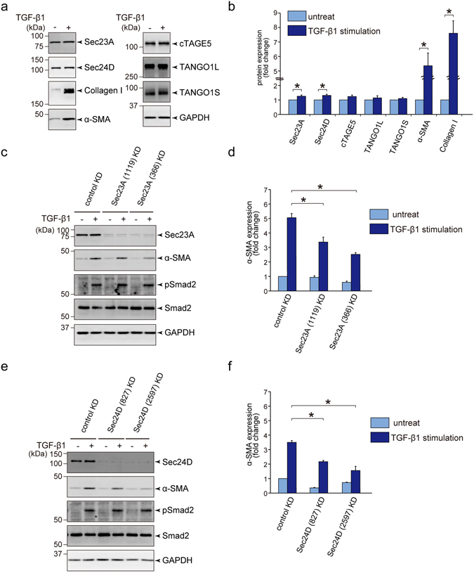
Figure 2.

Sec23A and Sec24D are required for activation of LX-2 cells upon TGF-β1 stimulation. (a and b) LX-2 cells starved for 24 h with DMEM supplemented with 0.5% FBS were untreated or treated with 1 ng/ml TGF-β1 and cultured for 3 days. Cells were extracted and subjected to SDS-PAGE followed by western blotting with anti-Sec23A, anti-Sec24D, anti-collagen I, anti-α-SMA, anti-cTAGE5, anti-TANGO1 and anti-GAPDH antibodies. (a) Representative immunoblots. (b) Quantification of immunoblots (n = 6). The band intensities were normalized to those of GAPDH. (c–f) LX-2 cells transfected with the indicated siRNA(s) were cultured for 24 h in DMEM supplemented with 10% FBS. After starvation for 24 h with DMEM supplemented with 0.5% FBS, the cells were untreated or treated with 1 ng/ml TGF-β1 and cultured for 3 days. Proteins were extracted and subjected to SDS-PAGE, followed by western blotting with anti-Sec23A (c), anti-Sec24D (e), anti-α-SMA, anti-Smad2, anti-pSmad2 and anti-GAPDH antibodies. (c,e) Representative immunoblots. (d,f) Quantification of immunoblots (n = 3). The band intensities of α-SMA were normalized to those of GAPDH. Error bars represent mean ± SEM. *P < 0.05. Uncropped original blots were shown in Fig. S3.
