Figure 4.

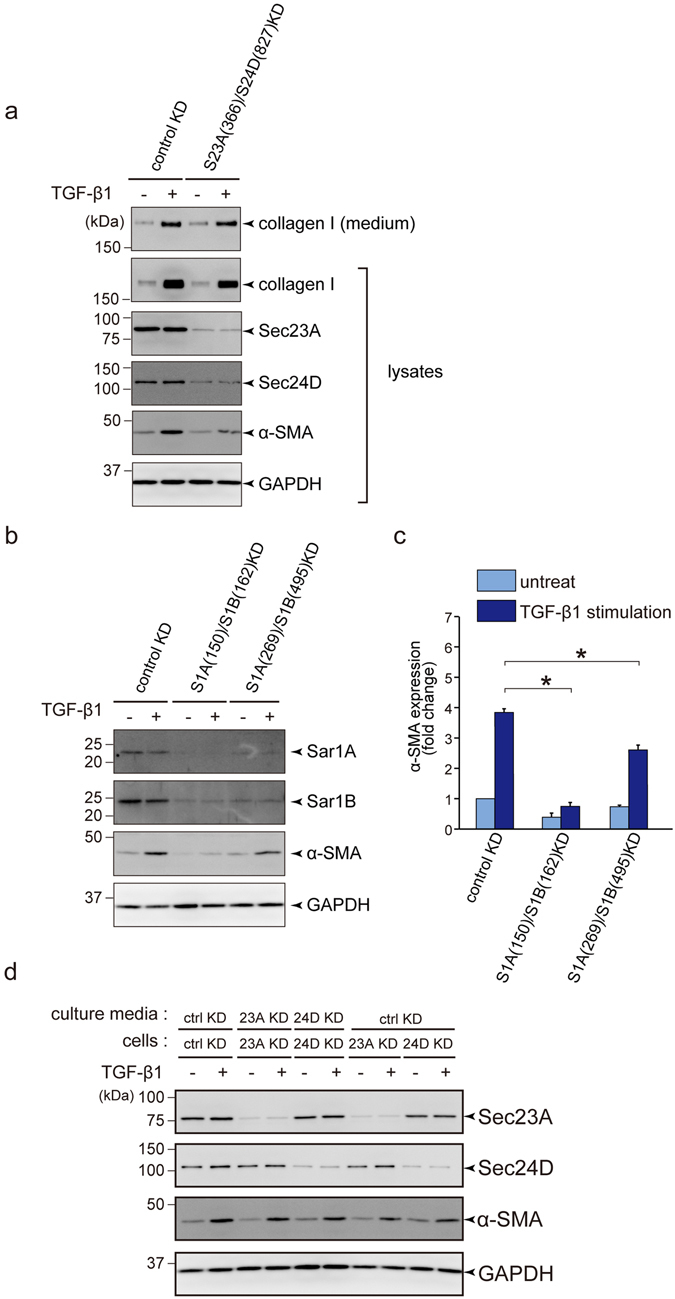
COPII-mediated ER to Golgi transport is required for activation of LX-2 cells upon TGF-β1 stimulation. (a) LX-2 cells transfected with the indicated siRNA(s) were cultured for 24 h in DMEM supplemented with 10% FBS. After starvation for 24 h with DMEM supplemented with 0.5% FBS, the cells were untreated or treated with 1 ng/ml TGF-β1 and cultured for 3 days. Proteins were extracted and subjected to SDS-PAGE, followed by western blotting with anti-collagen I, anti-Sec23A, anti-Sec24D, anti-α-SMA and anti-GAPDH antibodies (lysates). Medium was collected for SDS-PAGE followed by western blotting with anti-collagen I antibody (medium). Shown is a representative immunoblot analysis (n = 3). (b and c) LX-2 cells transfected with the indicated siRNA(s) were cultured for 24 h in DMEM supplemented with 10% FBS. After starvation for 24 h with DMEM supplemented with 0.5% FBS, the cells were untreated or treated with 1 ng/ml TGF-β1 and cultured for 3 days. Proteins were extracted and subjected to SDS-PAGE followed by western blotting with anti-Sar1A, anti-Sar1B, anti-α-SMA and anti-GAPDH antibodies. (b) Representative immunoblots. (c) Quantification of immunoblots (n = 3). The band intensities of α-SMA were normalized to those of GAPDH. (d) LX-2 cells transfected with the indicated siRNA(s) were cultured for 24 h in DMEM supplemented with 10% FBS. After starvation for 24 h with DMEM supplemented with 0.5% FBS, the cells were untreated or treated with 1 ng/ml TGF-β1 for 24 h. The culture medium was replaced with medium incubated with cells treated with the indicted siRNA(s) and further cultured for 48 h. Proteins were extracted and subjected to SDS-PAGE, followed by western blotting with anti-Sec23A, anti-Sec24D, anti-α-SMA and anti-GAPDH antibodies. Error bars represent mean ± SEM. *P < 0.05. Uncropped original blots were shown in Fig. S3.
