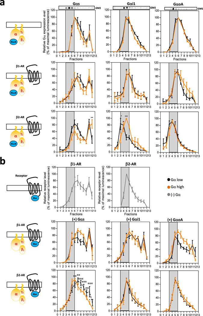
Figure 6.

Gα expression level influences Gα subunit and β-AR receptor compartmentalization in cholesterol-enriched membranes. Detergent-resistant-membranes were purified using a triton X-100 lysis method followed by a separation on sucrose gradient from (a) HEK293T cells co-expressing high (orange) and low (black) expression levels of Gαs-Rluc8 (left panel), Gαi1-Rluc8 (middle panel) or GαoA-Rluc8 (right panel) along with Gβ3 and Gγ2 subunits in the presence or not (upper panels) of untagged β-AR receptors, (b) HEK293T cells co-expressing β1-AR-Rluc or β2-AR-Rluc alone (upper panels) or in the presence of high (orange) or low (black) expression levels of untagged Gαs (left panels), Gαi1 (middle panels) or GαoA (right panels) and Gβ3 and Gγ2 subunits. Relative receptor or Gα subunit expression levels were quantified in each sucrose fractions by recording of the total luminescence. Results are expressed as the percentage of the maximal luminescence measured from all fractions in each experiment. Grey boxes highlight raft nano-domains enriched fractions, identified by detection of the GM1 protein (upper dot plot). Data represent the mean ± s.e.m. of at least three independent experiments. The statistical significance of the difference in membrane distribution in low versus high conditions (*) or versus receptor alone (¤) was assessed using two-way ANOVA followed by a Bonferroni posttest (*P < 0.05, **P < 0.01, ***P < 0.001).
