
Figure 5.
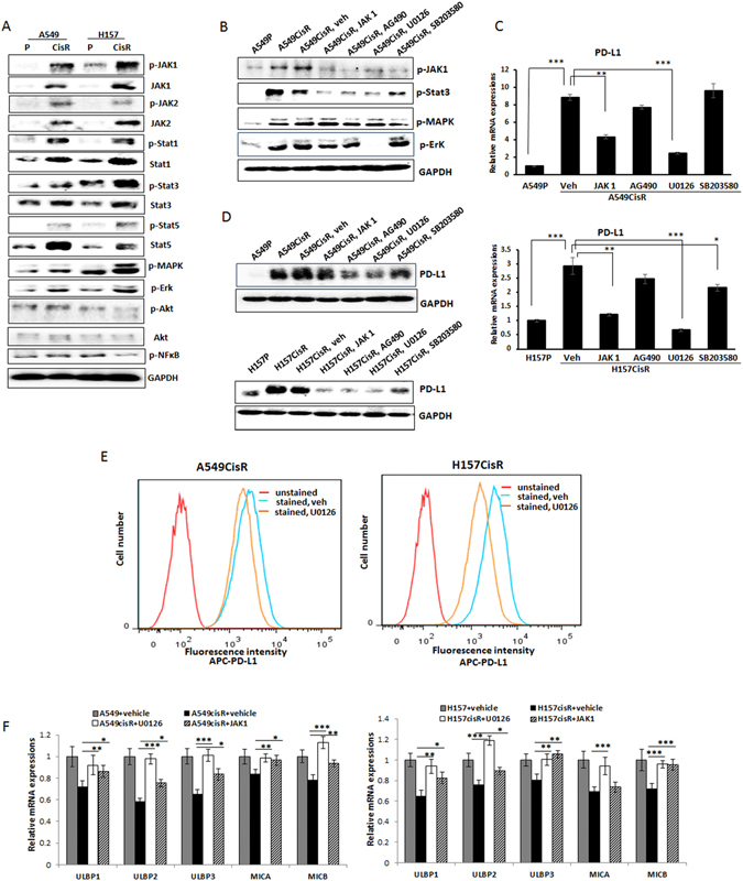
Effects of signaling pathway inhibition on PD-L1 levels in A549CisR and H157CisR cells. (A) Western blot analyses showing the expression/activation of several signaling molecules in parental and cisplatin-resistant cells. (B) Western blot analyses showing inhibition of each pathway in A549CisR cells upon inhibitor treatment. (C,D) PD-L1 levels in A549CisR and H157CisR cells upon treatment with inhibitors of indicated signaling pathways using qPCR analysis (C) and Western blot analysis (D). (E) Flow cytometric analyses of PD-L1 in cisplatin-resistant lung cancer cells. A549CisR and H157CisR cells were treated with U0126 (6 hours) (vehicle treated one as control) and positive stained APC-PD-L1 levels in these cells were analyzed. (F) NKG2D ligand expression on the A549CisR and H157CisR cells vs. parental cells. qPCR analyses show the recovery of NKG2D ligands upon addition of inhibitors of MEK/Erk and JAK signaling pathways. *p < 0.05, **p < 0.01, ***p < 0.001.