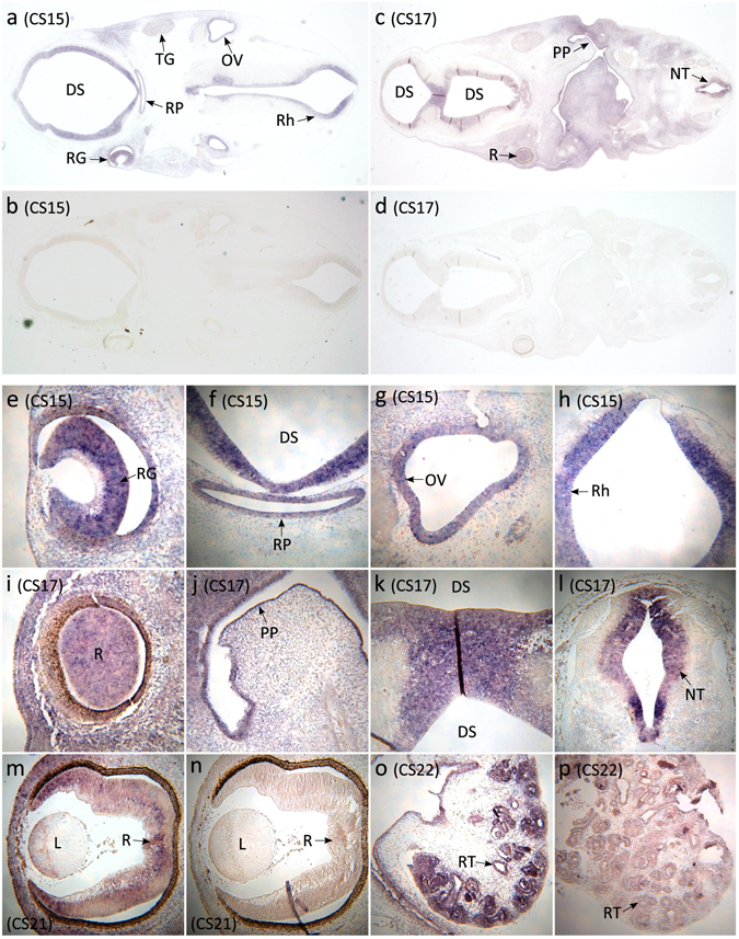
Figure 3.

YAP1 in situ hybridisation studies in the developing human. (a,b) In situ hybridisations of coronal sections of a CS15 human fetus using YAP1 antisense (a) and sense (negative control) (b) probes reveal expression in structures including the diencephalic superventricle (future third ventricle), retinal ganglion cell layer, Rathke’s pouch (future pituitary), trigeminal ganglion, otic vesicle, and rhombencephalon (future fourth ventricle). (c,d) In situ hybridisations of coronal sagittal sections of a CS17 human fetus using YAP1 antisense (c) and sense (negative control) (d) show expression of YAP1 in diencephalic superventricle, retina, primordium of the lateral palatine process, and neural tube. (e–h) High magnification images of YAP1 expression in the CS15 human fetus in multiple structures: (e) retinal ganglion cell layer, (f) diencephalic superventricle and Rathke’s pouch, (g) otic vesicle, and (h) rhombencephalon. (i–l) High magnification images of YAP1 expression in the CS17 human fetus in multiple structures: (i) retina, (j) primordium of the lateral palatine process, (k) diencephalic superventricle, and (l) the neural tube. (m,n) High magnification images of YAP1 expression in the retina of CS21 human fetus: (m) antisense, and (n) sense. (o,p) High magnification images of YAP1 expression in the kidney of CS22 human fetus: (o) antisense, and (p) sense. Abbreviations: DS = diencephalic superventricle; L = lens; NT = neural tube; OV = otic vesicle; PP = primordium of the lateral palatine process; R = retina; RG = retinal ganglion cell layer; Rh = rhombencephalon; RP = Rathke’s pouch; RT = renal tubules; TG = trigeminal ganglion.
