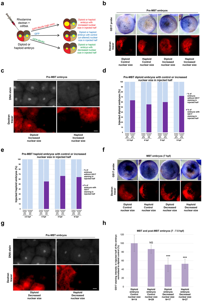
Figure 2.

GS17 in situ hybridization in embryos with altered nuclear size and ploidy (a) One blastomere of a two-cell haploid or diploid embryo was coinjected with rhodamine-labeled dextran as a tracer and mRNA to alter nuclear size in half of the embryo. For control embryos, 3.5, 4, 5, 6, and 7 hpf correlate to stage 6, 6.5, 7, early 8, and 8, respectively. (b) GS17 in situ hybridization was performed on pre-MBT embryos with increased nuclear size in half of the embryo. Bright-field images of embryos stained for GS17 (purple) are shown in the top panels. The corresponding rhodamine (red) fluorescence images indicating cells in the embryo that received the microinjected mRNA are shown in the bottom panels. Representative embryos are shown. (c) 5 hpf diploid and haploid embryos with increased nuclear size in the injected halves were stained with Sytox Green and cleared for imaging. The top panels are fluorescent images of embryos stained for DNA. The bottom panels are the corresponding rhodamine fluorescence images indicating cells in the embryo that received the microinjected mRNA. Representative embryos are shown. Scale bar, 20 µm. (d,e) The graphs show the percentage of pre-MBT diploid (d) and haploid (e) embryos with (dark purple) or without (light blue) differential GS17 staining. Embryos were scored as showing differential GS17 staining as long as some cells that received the microinjected mRNA stained positively for GS17. N = number of embryos. (f) 7 hpf embryos with decreased nuclear size in half of the embryo were subjected to GS17 in situ hybridization. Imaging was performed as in (b). (g) 5 hpf diploid and haploid embryos with decreased nuclear size in the injected halves were stained with Sytox Green and cleared. Imaging was performed as in (c). Scale bar, 20 µm. (h) The graph shows GS17 staining intensity in injected halves of 7-7.5 hpf diploid and haploid embryos with control or decreased nuclear size, normalized to the diploid control. N = number of embryos. Error bars represent SD. ***p < 0.001; NS = not significant.
