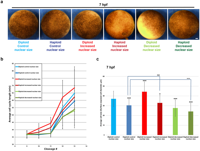
Figure 4.

Cell cycle timing in embryos with altered nuclear size and ploidy. One-cell diploid and haploid embryos were microinjected with mRNA to alter nuclear size, and bright-field time-lapse imaging was performed on the animal pole surface at 5-min intervals at 21 °C. (a) Still frame images of 7 hpf embryos. Scale bar, 100 µm. (b) Cell-cycle lengths were measured for at least five cells per embryo starting at the ninth cell division for three embryos per condition. Data from two independent experiments are shown; error bars represent SD. (c) Cell sizes were estimated by quantifying the diameter of surface-exposed cells on the animal pole of 7 hpf embryos. For each embryo, several random regions were selected for cell size quantification. Cells from at least two embryos were quantified for each condition. Total number of cells quantified: diploid control, n = 34; haploid control, n = 34; diploid increased nuclear size, n = 42; haploid increased nuclear size, n = 43; diploid decreased nuclear size, n = 22; haploid decreased nuclear size, n = 36. Error bars represent SD. ***p < 0.001; *p < 0.05; NS = not significant.
