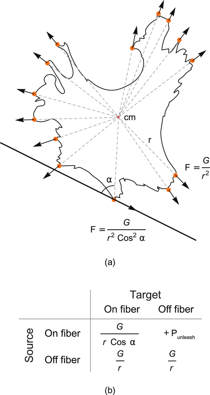
Figure 7.

Schematic representation of cell spreading and cell–nanofibre interaction. (a) The cell shape was taken from the experimental database. All protrusions are marked with orange circles. Arrows show the direction of the resulting force driving the cell expansion at the attachment sites. If no guiding fibres exist, the cell spreads equidirectionally, and the forces are directed from the centre of mass of the cell (cm). If a nanofibre exists (at the bottom), and the cell is already attached to it, then the force is aligned with the fibre. (b) Energy terms, applied for different copy attemts of the attachment site to the non-attachment subcell, from the isotropic sibstrate to the fiber or in opposite direction.
