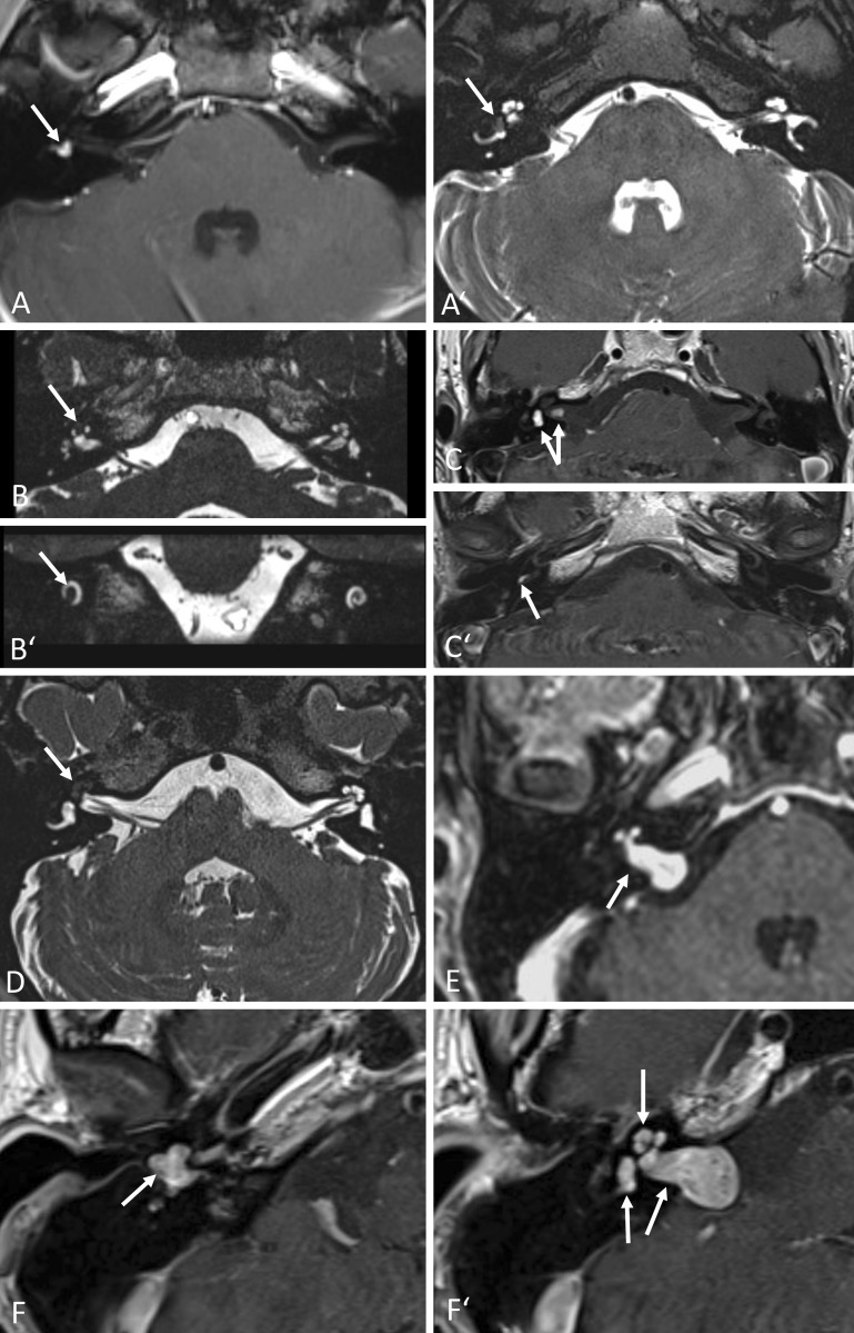
Fig. 1.
Intralabyrinthine schwannomas (ILS); MRI findings (arrows). A, A′ Typical contrast enhancement on T1-w (A) and “filling defect” on T2-w (A′) images of histologically confirmed intravestibular schwannoma (patient 8). Axial images. B, B′ Intracochlear schwannoma in the apical and partially in the middle turn (T2-w, B axial, B′ coronal); not histologically confirmed (patient 4). C, C′ Three locally separated schwannomas: intravestibular, intrameatal (C), basal cochlear turn (C′); not histologically confirmed (T1-w, c.m., axial; patient 6). D Intracochlear schwannoma of the entire cochlea, histologically confirmed (T2-w, axial; patient 9). E Tumor probably originating from the internal auditory canal (IAC) with extension into the cerebellopontine angle and transmodiolar into the basal and middle cochlear turn; histologically confirmed (T1-w, c.m., axial; patient 12). F, F′ Transotic ILS with extension from the tympanic membrane via the entire labyrinth and the IAC to the cerebellopontine angle; histologically confirmed (T1-w, c.m., axial; patient 10). w Weighted, c.m. contrast medium

