
Figure 6.
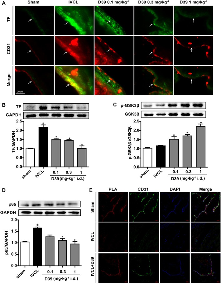
D39 suppressed endothelial TF expression by modulating the GSK3β/p65 signalling pathways through inhibiting the dissociation of NMMHC IIA from TNFR2 in vivo. (A) IVC frozen sections were prepared, and TF expression was analysed by immunofluorescence. (B) IVC lysates were prepared, and TF expression was analysed by Western blots. (C, D) IVC lysates were prepared, and p‐GSK3β, GSK3β and p65 expressions were analysed by Western blots. (E) IVC frozen sections were prepared and used for PLA to confirm NMMHC IIA–TNFR2 interaction (red signal), CD31 staining (green) and DAPI staining (blue). Data shown are the means ± SEM of five mice per group. # P < 0.05, significantly different from the sham group; *P < 0.05, significantly different from the IVCL group.