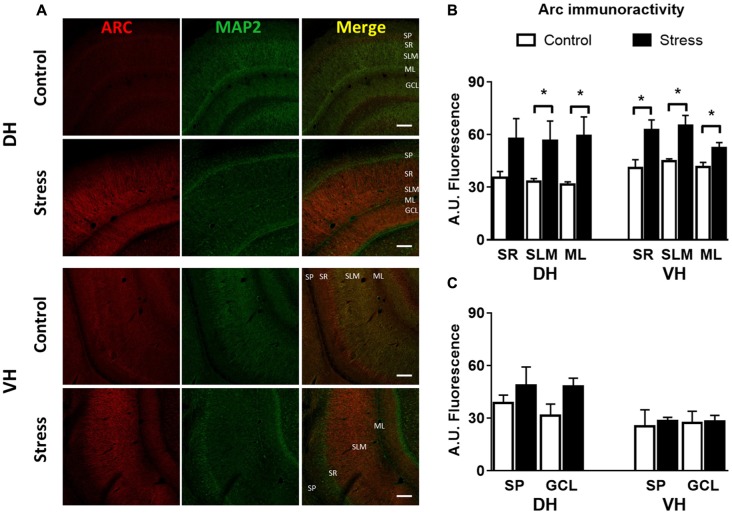
Figure 4.
Chronic stress produces an enhancement in Arc immunoreactivity at synaptic layers of DH and VH. (A) Confocal microscopic images displaying immunoreactivity of Arc (red) and MAP2a (green) in DH and VH of control and chronically stressed animals. Scale bar = 200 μm. Quantification of the immunoreactivity detected at the dendritic (B) and somatic (C) layers of both DH and VH. SP, stratum pyramdalis; SR, stratum radiatum; SLM, stratum lacunosum moleculare; ML, molecular layer; GCL, granular cell layer. Graphs represent mean ± SEM of each group. Differences between control and stressed animals were determined by two-tail Mann-Whitney test. *P < 0.05.

