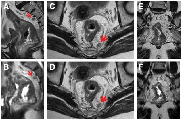
Figure 1.

Oblique axial, oblique coronal and sagittal section of 2D T2-weighted FSE (A, C, E) and 3D CUBE (B, D, F) in a 61-year-old woman with rectal cancer after neoadjuvant therapy. The tumor of rectal wall and the lymph node in the mesorectum were well shown on multiple planes of 3D CUBE and 2D (the lymph node being represented by arrow). The mild invasion into mesorectal fat implied T3 stage, which was confirmed pathologically.
