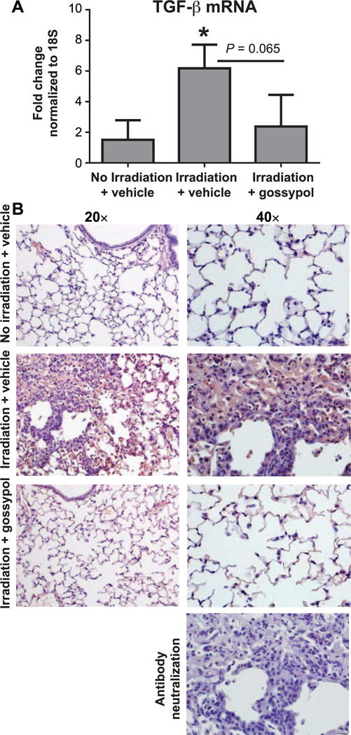
FIG. 3.

Gossypol dampens radiation-induced TGF-β expression and activation. Mice were irradiated and vehicle or gossypol treated as indicated in Materials and Methods. Whole lung tissue TGF-β mRNA was quantified using RT-PCR. Data is expressed as fold change compared to nonirradiated controls, normalized to 18S (panel A). Data are shown as mean ± SEM for n = 5–8 mice. *P ≤ 0.05 compared to nonirradiated vehicle-treated controls (ANOVA). Panel B: Lung tissue sections were stained with an antibody to active TGF-β (red) and counterstained with hematoxylin (blue) as described in Materials and Methods. One representative mouse is shown from each treatment group. For negative control, antibody neutralized with active human recombinant TGF-β was used at 30 min prior to staining irradiated lung tissue. Images were taken at 20× and 40× magnification. Scale bars represent 100 μm at 20× and 50 μm at 40×.
