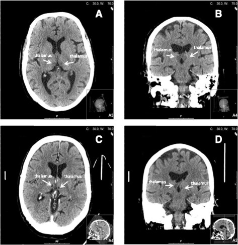
Fig. 1.

a Head computed tomography (axial) performed on the day of admission was initially assessed as normal, although signs of bilateral ischemia in the thalami actually were visible. b Head computed tomography (coronal) performed on the day of admission was initially assessed as normal, but later re-evaluated to be bilateral ischemia in the thalami. c Head computed tomography (axial) performed on day 24 of hospitalization showed bilateral ischemia in the medial areas of the thalami. d Head computed tomography (coronal) performed on day 24 of hospitalization showed bilateral ischemia in the medial areas of the thalami
