Figure 3.

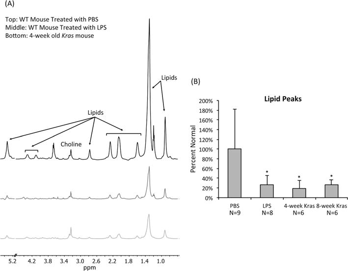
Neoplasia is associated with a drop in lipid levels in mouse tissue samples. (A) Representative pulse–acquire spectra of pancreata from a wild type mouse treated with PBS, a wild type mouse treated with LPS, and a 4 week old Kras mouse. Spectra display a dramatic drop in lipid levels associated with LPS treatment and Kras. All spectra illustrate normalization to sample weight. The peaks at 1.20 and 3.67 ppm are due to the use of ethanol during the tissue collection procedure. The 4.60–5.15 ppm region was removed to exclude the residual water signal. (B) Average levels of lipids present at 1.30 ppm as determined from 1H HR-MAS of pancreatic tissue samples of PBS-treated wild type mice, LPS-treated wild type mice, 4 week old Kras mice, and 8 week old Kras mice. Lipid levels are lower in LPS-treated wild type, 4 week old Kras, and 8 week old Kras mice compared with PBS-treated wild type mice. *p < 0.05.
