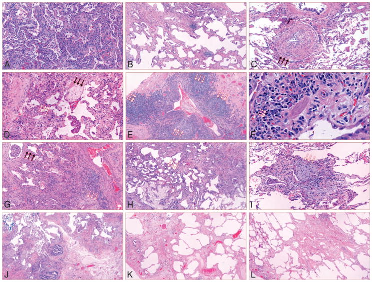
Figure 3.
Histopathologic features of samples from patients with idiopathic interstitial pneumonia. A, Cellular nonspecific interstitial pneumonia (NSIP). B, Fibrotic nonspecific interstitial pneumonia NSIP. C, Pulmonary vasculopathy in specimen with NSIP. Pulmonary vasculopathy (black arrows). D, Overlap of NSIP with organizing pneumonia (OP): NSIP, white arrows; OP, black arrows. Multiple foci of OP were present in the tissue specimen. E, Interstitial lymphoid aggregates with germinal centers (white arrows). F, Lymphoplasmacytic infiltration with lymphoid follicles (white arrows). G, Usual interstitial pneumonia with NSIP; NSIP in upper left (black arrows); fibroblastic focus in bottom right (white arrows). H, Usual interstitial pneumonia with fibrosing NSIP (fibrosing NSIP, white arrows). I, Usual interstitial pneumonia with OP (white arrows). Multiple foci of OP were present in tissue specimen. J, Usual interstitial pneumonia only. K and L, Unclassifiable interstitial pneumonia. A through I, The idiopathic interstitial pneumonia meets interstitial pneumonia with autoimmune features criteria. J through L, The IIP does not meet IPAF criteria (hematoxylineosin, original magnifications ×100 [A, C, D, and G], ×40 [B, E, H, and J–L], × 600 [F], and × 200 [I]).

