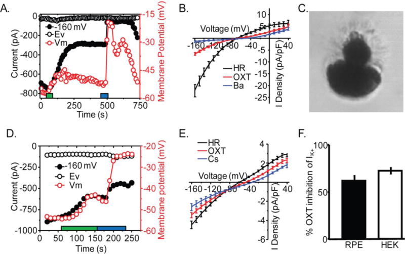
Fig. 7.

OXT activation of OXTR inhibits Kir7.1 channel. A) Representative HEK-OXTR whole-cell current time course measured at cell potentials −68mV (Ev) and −160 mV during application of a ramp voltage protocol from −160 to −40 mV every 10 second while holding the cell at −10 mV. In addition, the membrane potential of the cell (Vm) is plotted over the time course of the experiment. The duration of OXT (red) and Kir7.1 blocker Cs2+ (blue) application is indicated. B) Average plot of I-V whole cell current at baseline (Black; n=9), and after application of OXT (Red; n=9), Cs2+ (Blue; n=9). C) Image of dissociated mouse RPE cell with polarized morphology representative of cells selected for recording. D) Representative mouse RPE whole-cell current time course measured at cell potentials −62 (Ev), and −160 mV during application of the same ramp protocol as in A. The membrane potential of the cell is plotted over the time course of the experiment. Duration of application of OXT (red) and Cs2+ (blue) is indicated. The duration of OXT (red) and Kir7.1 blocker Cs2+(blue) application is indicated. E) Average plot of I-V whole cell current before (Black; n=7), and after application of OXT (Red; n=7) or Cs2+ (Blue; n=3). F) Comparison of ratio of Cs2+ sensitive current during OXT treatment relative to current in HR in murine RPE and HEK-OXTR cells. Data is from at least three independent experiments and represented as mean ± SEM.
