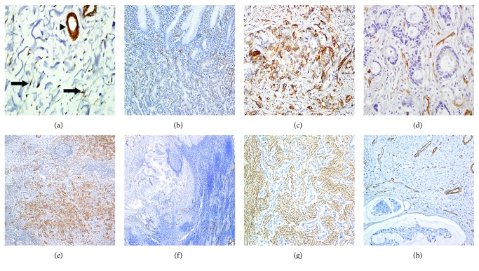
Figure 1.
Representative caveolin-1 (Cav-1) expression. In nonneoplastic gastric tissue, Cav-1 expression was detected in fibroblast (arrow) and vessel walls (arrow head) in stroma (a), while Cav-1 showed a scant expression in parietal cells in epithelium (b). Tumor cells showed high (c) and low expression (d) of Cav-1 in the stomach and high (e) and low expression (f) of metastatic lymph node. Stromal Cav-1 immunoreactivity was observed in low (g) and high (h).

