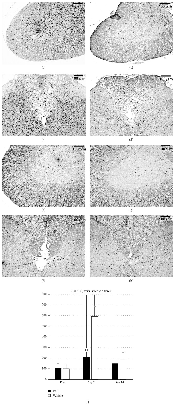
Figure 5.
Oral administration of RGE suppressed aggregation of Iba-1-positive microglial cells in injured spinal cord at 7 days after SCI in rats. (a–h) Representative photomicrographs of Iba-1 immunostaining in sections from damaged spinal cord of rats at 7 and 14 days after SCI ((a)–(d): 7 days after SCI; (e)–(h): 14 days after SCI; (a), (b), (e), and (f): vehicle; (c), (d), (g), and (h): RGE (350 mg/kg/day)). Scale bar = 100 μm. (i) Relative optical density (ROD) as % of Iba-1 immunoreactivity in injured spinal cord. There was a significant decrease in Iba-1 immunoreactivity at 7 days after SCI in RGE-treated rats (n = 5 in each group). All values are presented as mean ± SD. ∗∗(p < 0.01) indicates significantly lower values than vehicle-treated control.

