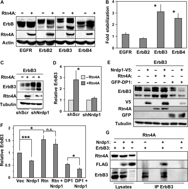
Fig. 2. Rtn4A interferes with Nrdp1-mediated ErbB3 degradation.
(A) HEK293T cells were cotransfected with each of the human ErbB family members shown at the bottom of the blot along with either empty vector (−) or vector containing Rtn4A (+). Lysates were blotted for ErbB receptors, Rtn4A, and actin. (B) Five independent experiments such as that illustrated in (A) were quantified, and the fold accumulation of each ErbB receptor in the presence of Rtn4A relative to its abundance in the absence of Rtn4A was plotted. (C) Cells were cotransfected with ErbB3 along with either empty vector or vector containing Rtn4A, plus either scrambled control shRNA (shScr) or the Nrdp1-directed shRNA KD1 (13), as indicated. Lysates were blotted with antibodies recognizing ErbB3, Rtn4A, and tubulin. (D) Eight independent experiments such as that illustrated in (C) were quantified, and the fold change in ErbB3 abundance in response to the various conditions was plotted. (E) HEK293T cells were cotransfected with ErbB3 along with Rtn4A, Nrdp1-V5, or GFP-DP1, as indicated, and lysates were blotted with antibodies recognizing ErbB3, V5, Rtn4A, GFP, and tubulin. (F) Three independent experiments such as that illustrated in (E) were quantified, and the relative effects of the various conditions on ErbB3 abundance were plotted. (G) HEK293T cells were co-transfected with Rtn4A together with Nrdp1-FLAG, ErbB3, or both, as indicated. Lysates (left lanes) from cells treated overnight with 1.5 µM MG132 were immunoprecipitated with antibodies recognizing ErbB3 (right lanes), and lysates and precipitates were blotted with antibodies recognizing Rtn4A, FLAG, and ErbB3. Data are representative of three independent experiments. *P < 0.05; ***P < 5 × 10⁻⁵; n.s., not significant by Student’s t test.

