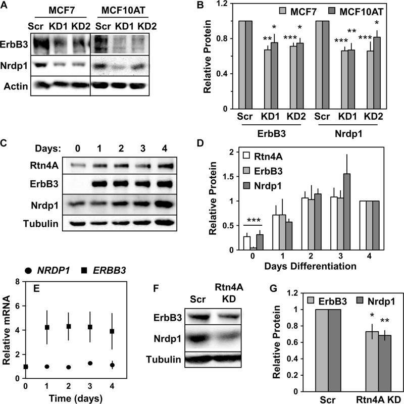
Fig. 3. Rtn4A depletion destabilizes endogenous ErbB3 and Nrdp1.
(A) MCF7 (left lanes) and MCF10AT (right lanes) cells were treated with either scrambled control siRNA (Scr) or siRNA targeting Rtn4A (KD1 and KD2). Lysates were immunoblotted to show ErbB3, Nrdp1, and actin. (B) Four independent experiments such as that illustrated in (A) were quantified, and the fold change in ErbB3 and Nrdp1 protein abundance was plotted for each siRNA. (C) C2C12 myotubes were differentiated for 4 days. Lysates were immunoblotted to show Rtn4A, ErbB3, Nrdp1, and tubulin. (D) Four independent experiments such as that illustrated in (C) were quantified, and the fold change in the abundance of Rtn4A, ErbB3, and Nrdp1 proteins from day 0 to 4 was plotted. (E) NRDP1 and ERBB3 transcript abundance in three independent experiments was determined by qRT-PCR in differentiating C2C12 myotubes. (F) Lysates from C2C12 myoblasts treated with scrambled control siRNA or siRNA targeting mouse Rtn4A (Rtn4A KD) before differentiation were immunoblotted for Nrdp1, ErbB3, and tubulin. (G) Six independent experiments such as that illustrated in (F) were quantified, and the fold change in Nrdp1 and ErbB3 protein abundance was plotted. *P < 0.05; **P < 5 × 10⁻³; ***P < 5 × 10⁻⁵ by Student’s t test.

