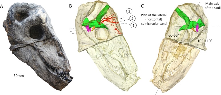
Figure 1. The skull of Moschops capensis AM4950 in lateral view.
(A) Photograph of the skull. (B) Reconstruction of the skull (right side, bone transparent) to reveal the neural structures discussed in this paper. (C) Reconstruction of the skull (left side, bone transparent) showing the endocast, bony labyrinth and the angle between the plane of the lateral semicircular canal and the main axis of the skull. Numbers indicate the position of the cross sections in the subsequent figures. EmV, emissary veins; End, endocranial cast; Hyp, hypophyseal fossa; Lab, bony labyrinths; Pin, pineal tube. Photo by LN.

