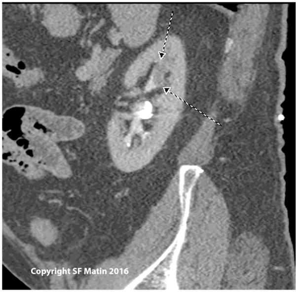
Figure 1.
Sagittal view of a computed tomography scan showing a filling defect representing a soft tissue mass in the upper pole of the left collecting system, in a patient with prior endoscopic therapy for low grade left ureteral tumors. Biopsy in this case showed high grade papillary tumor. Nephroureterectomy showed parenchymal invasion (stage pT3).

