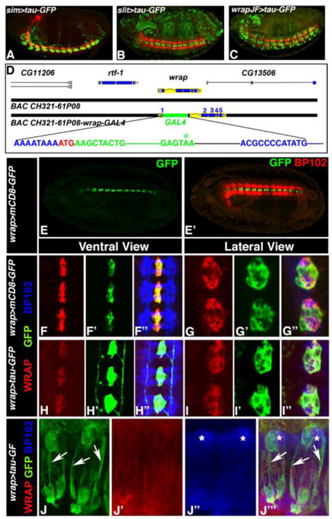
Figure 1. Generation and expression of wrapper-GAL4.

(A-C) Confocal images of Stage 16 embryos of sim-GAL4/UAS-tauGFP (A), slit-GAL4/UAS-tauGFP (B) and wrapper-GAL4/UAS-tauGFP (C, Janelia GAL4 stock abbreviated as JF) show expression of GFP (green) and BP102 (red). (D) Design of the wrapper-GAL4 construct using BAC CH321-61P08. The DNA sequence in blue is from the wrapper gene with its initiator codon ATG shown in red. The sequence in green is the GAL4 sequence and the termination codon is shown in green with asterisk. Further downstream the wrapper sequence continues in blue. No wrapper sequences were deleted but only disrupted with inframe insertion of GAL4. (E) Confocal image of Stage 16 embryo of wrapper-GAL4/UAS-mCD8-GFP in ventral view shows GFP expression in MG (E) and BP102 in CNS axons (E′). (F-F″ and G-G″) Higher magnification confocal images of Stage 16 embryos of wrapper-GAL4/UAS-mCD8-GFP (F-F″, ventral view, and G-G″ lateral view) shows an overlap of Wrapper with GFP (F″ and G″). (H-H″ and I-I″) Confocal images of Stage 16 wrapper-GAL4/UAS-tau-GFP embryos in ventral view (H-H″) and lateral view (I-I′) also display an overlap of Wrapper (H and I) with GFP (H′ and I′) at the MG. GFP expression can also be seen in glial processes (green asterisk, H′). BP102 (blue) was used for labeling CNS axonal scaffold. Embryos in A, B and C show a lateral view and are oriented anterior to the left and dorsal top. Embryo in E shows a sagittal view with anterior to the left. Higher magnification images in F-I″ have anterior to the top. (J-J‴) Confocal images of MG and their ensheathing processes in third instar ventral nerve cord of wrapper-Gal4/UAS-tau-GFP labeled with GFP (J, J‴, arrows), Wrapper (J′, J‴) and BP102 (J″, J‴, asterisks).
