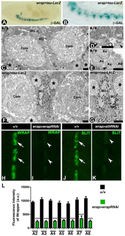
Figure 2. Utility of wrapper-GAL4 in the study of CNS midline.

(A, B) β-Gal staining of wrapper-GAL4/UAS-tau-lacZ stage 16 embryo show lacZ expression specifically in the MG at a low magnification (A) and high magnification (B). (C-E) Electron micrographs of embryonic stage 16 wild type midline displayed commissural axon tracts (Com) separated by MG (C). Also visible are the neuronal cell bodies in the CNS (asterisk). Higher magnification images shown with MG and their processes (arrowheads, D) and axons (Ax, E) surrounded by MG processes (arrowhead). (F, G) Electron micrographs of stage 16 wrapper-GAL4/UAS-tau-lacZ reveal MG cells and processes decorated by β-Gal reaction product crystals (F, G). Note the commissural axon tracts crossing the midline are surrounded by MG processes and are highlighted by β-Gal reaction product crystals (G). (H-L) Confocal images of stage 16 embryonic midline of wrapper-GAL4/UAS-wrapper-RNAi (I) and wrapper-GAL4/UAS-slit-RNAi (K) show a reduction in the levels of Wrapper (I, quantified in L) and Slit (K) compared to their wild type counterparts (H and J, respectively). Scale bars: C, F= 5μm; D, E= 0.2 μm; G= 2 μm. Anterior is to the left in A, B and to the top in H-K. a.u.= arbitrary units
