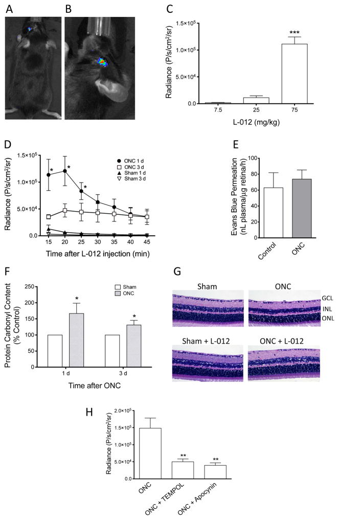
Figure 1. Characterization of L-012 chemiluminescence in mouse optic nerve crush-induced retina injury.
(A) L-012 chemiluminescence was detected in the injured (left) mouse eyes following ONC. No chemiluminescence in the contralateral (right) sham control eye. (B) For subsequent quantifications, animals were positioned so that the injured eye was facing the camera. Radiance is shown as pseudocolor overlay with photo image of the mouse head. (C) Relationship of L-012 dose and radiance at 20 min after L-012 injection, 1 d post ONC. ***: P<0.001 vs the 7.5 mg/kg group by one-way ANOVA and Bonferroni’s test (n=5). (D) Time courses of L-012 chemiluminescence. Quantification of radiance was obtained from 15 min to 45 min, in 5-min exposure intervals, after L-012 injection (75 mg/kg, ip) at 1 d and 3 d after ONC. *: P<0.05 vs the respective sham control groups (n=5) by unpaired Student’s t-test. The data points 3 d after ONC were not statistically different. (E) Evaluation of blood-retina barrier function by Evans blue retina permeation assay. At 1 d after ONC, mice were injected with Evans blue intravenously. Levels of Evans blue in retinas in the ONC-injured and its contralateral sham control eyes were assayed and compared 2 h later. No statistically significant difference (P>0.05) was detected by paired Student’s t-test (n=4). (F) Quantification of ONC induced retinal oxidative damage at 1 d and 3 d post-crush by protein carbonyl ELISA. *: P<0.05 vs the respective sham control groups (n=6) by unpaired Student’s t-test. (G) Histological evaluation of retinal cross sections of sham or ONC mice, with or without multiple L-012 injections (75 mg/kg, ip, at 1 d, 3 d, 5 d, and 7 d post-crush). Mice were euthanized and retina samples collected at 7 d after sham or ONC operation. Other than a reduced cell density in the ganglion cell layer in the ONC groups, no additional abnormality was observed. L-012 did not appear to damage the retina. Three animals from each group were assessed. GCL: ganglion cell layer, INL: inner nuclear layer, ONL: outer nuclear layer. (H) Effect of TEMPOL (100 mg/kg, ip, 15 min prior to L-012 injection) and apocynin (50 mg/kg, ip, 15 min prior to L-012 injection) on the L-012 (75 mg/kg, ip) signal at 1 d post-ONC. Radiance values at 20 min after L-012 injection are shown. **: P<0.01 vs the ONC group by one-way ANOVA and Bonferroni’s test (n=5). All data are shown as mean ± SEM.

