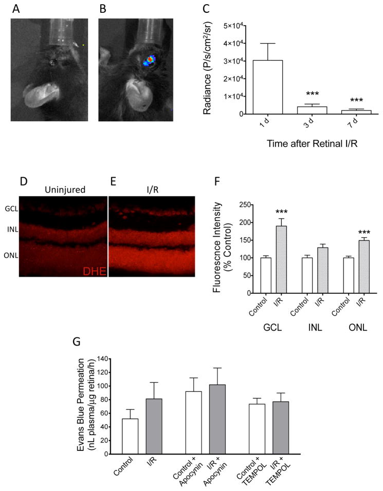
Figure 2. Retinal accumulation of ROS after ischemia/reperfusion.
(A) No chemiluminescence in the contralateral control eye. (B) L-012 chemiluminescence was detected in the injured (left) mouse eyes following retinal I/R. (C) Chemiluminescence signal at 20 min after L-012 injection (75 mg/kg, ip) at indicated time points after retinal I/R. ***: P<0.001 vs the 1 d group by one-way ANOVA and Bonferroni’s test (n=7). (D, E, F) Post mortem detection of superoxide in retina with DHE. Representative images of retina cross sections from control (D) and I/R (E) groups labeled with DHE. Retina samples were collected at 24 h after I/R. (F) Differences in fluorescence intensities were quantified in the three nucleated layers of the retina. Superoxide was observed to be significantly increased in the ganglion cell layer (GCL) and outer nuclear layer (ONL), but not the inner nuclear layer (INL). ***: P<0.001 vs control by unpaired Student’s t-test (n=4). Data are shown as mean ± SEM. (G) Evaluation of blood-retina barrier function by Evans blue retina permeation assay. At 1 d after I/R, mice were injected with Evans blue intravenously. In groups with apocynin (50 mg/kg) or TEMPOL (100 mg/kg) treatments, the drugs were administered intraperitoneally 15 min prior to Evans blue injection. Levels of Evans blue in retinas of the I/R-injured and contralateral control eyes were assayed 2 h after Evans blue injection. No statistically significant difference (P>0.05) was detected by paired Student’s t-test (No drug treatment groups: n=10; Apocynin-treated groups, n=6; TEMPOL-treated groups, n=7).

