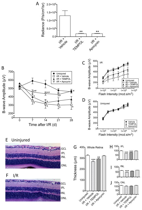
Figure 3. Protective effects of TEMPOL and apocynin against retina ischemia/reperfusion-induced damge.
(A) L-012 (75 mg/kg, ip) chemiluminescence was significantly reduced by TEMPOL (100 mg/kg, ip, 15 min prior to L-012 injection) and apocynin (50 mg/kg, ip, 15 min prior to L-012 injection) at 1 d post-injury. Radiance values at 20 min after L-012 injection are shown. **: P<0.01 vs the “I/R + Vehicle” group by one-way ANOVA and Bonferroni’s test (n=5). (B) Reduction of superoxide was protective against I/R-induced loss of visual function. Scotopic flash ERG was measured weekly following retinal I/R. The mice were treated with either vehicle, TEMPOL (100 mg/kg, ip), or apocynin (50 mg/kg, ip) 30 min prior to injury, then once daily from 1 d to 7 d. The time course plot shows b-wave amplitudes in response to a flash intensity of 3,000 mcd.s/m2 in each treatment group. While all three I/R groups displayed signifcant loss of b-wave amplitudes compared to the uninjured control group as early as 7 d, apocynin produced significant protection compared to the “I/R + Vehicle” group. TEMPOL treatment demonstrated a trend of protection, but the improvement was not significantly different from the “I/R + Vehicle” group. *: P<0.05 vs “Uninjured” group, and #: P<0.05 vs “I/R + Vehicle” group, by one-way ANOVA followed by Bonferroni post hoc test (n=6-7). (C) b-wave amplitudes in response to all flash intensities of the same animals in Figure 3B at 28 d post-injury. Amplitudes of apocynin-treated mice were similar to the uninjured eyes, while TEMPOL produced a trend of improvement compared to the “I/R + Vehicle” group. (D) TEMPOL and apocynin did not affect b-wave amplitudes of the contralateral uninjured eyes. At 28 d, the mice were euthanized and their retinas sectioned and H&E stained. L-012 did not affect the gross morphology of the mouse retina (E) & (F). However, I/R significantly decreased thicknesses of the whole retina (G), inner plexiform layer (IPL)(H), and inner nuclear layer (INL)(I), but not outer nuclear layer (ONL)(J). Apocynin was significantly protective against retinal thinning (G–J). It appeared that retina layers of the TEMPOL-treated mice were thicker than those of the “I/R + Vehicle” group, the differences were typically not significant (G–J). **: P<0.01, ***: P<0.001 vs Uninjured group by one-way ANOVA followed by Bonferroni post hoc test (n=6–7). Data are shown as mean ± SEM.

