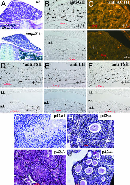
Fig. 5.
Histology and immunocytochemistry of pituitary. (A) Hematoxylin/eosin staining of sagittal sections of the pituitary of 8-wk-old male wt and smpd3–/– mice, ×120. Immunocytochemistry of paraffin-embedded sagittal sections of the pituitary of wt and smpd3–/– mice (p20) is shown. Antibodies stained only their specific peptide hormone antigen in the anterior pituitary lobe. (B–F) Primary antibody: anti-GH (B); anti-ACTH (C); anti-follicle-stimulating hormone (FSH) (D); anti-luteinizing hormone (E); anti-TSH (F). (B–D and F) Secondary antibodies: horseradish peroxidase-conjugated anti-rabbit IgG (B, D and F); Alexa-conjugated anti-rabbit IgG (C). a.l., anterior lobe; i.l., intermediate lobe. (Upper) Control +/+.(Lower) smpd3–/–. Light microscopy of cross sections: hematoxylin/eosin-stained testis in p42 wt (G) and p42 smpd3–/– (I) male mouse; p42 ovary of control wt (H) and smpd3–/– (J) female mouse.

