
Figure 3.
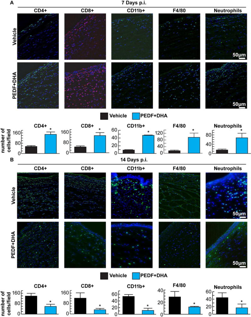
Rabbit corneas were stained with CD4+, CD8+, CD11b+, F4/80 and anti-neutrophil antibodies. The representative images show inflammatory cells in corneal sections at 7 (A) and 14 (B) days p.i. DAPI was used to stain the nuclei (blue). Positive cells from six corneal sections from each different cornea/condition were counted in five to six randomly selected fields in a blind fashion. Values are average of six corneas ± SD. The experiment was done four times with similar results. * Significant differences were analyzed by Student’s t-test (p<0.05).