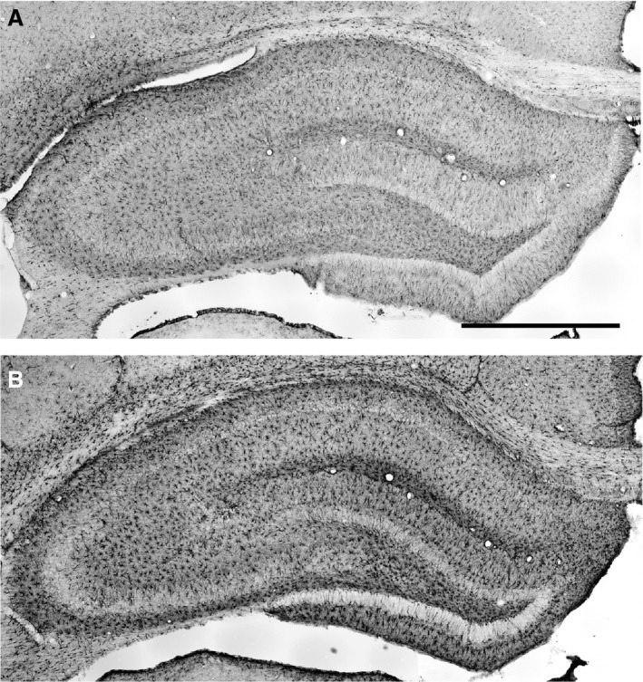
Figure 1.

Frontal sections of the hippocampal formation immunostained for glial fibrillary acidic protein (GFAP) to visualize astrocytes (Bregma − 3.8 mm, Paxinos & Watson, 1986). The two examples showing the range of differences in GFAP immunostaining are taken from rats given LPS on P06 which were not subjected to seizure induction (A) and from rats given LPS on P30 and experiencing seizures induced in adulthood (B). Scale bar: 1 mm.
