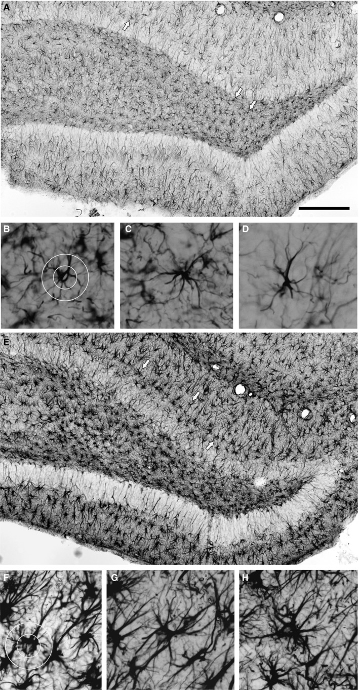
Figure 4.

Assessment of astrocyte morphology in the DG area. (A, E) Enlarged segments of Fig. 1A and B taken, respectively, from rats given LPS on P06 which were not subjected to seizure induction and from rats given LPS on P30 and experiencing seizures induced in adulthood. Scale bar: 500 μm. Examples of GFAP‐immunopositive astrocytes indicated by arrows in (A) and (E) are shown in (B–D) and (F‐H), respectively. The degree of ramification of astrocyte processes (branching index) was defined according to Garcia‐Segura & Perez‐Marquez (2014). Two circles of 25 and 50 μm diameter (B and F) were centered on the astrocyte cell body and intersections between the circles; the astrocyte processes were counted, and the ratio between number of intersections with the outer and inner circles was calculated.
