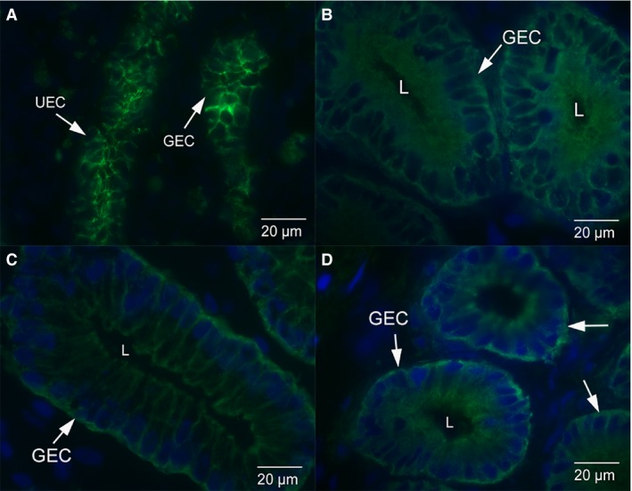
Figure 2.

Immunofluorescence micrographs of uterine glandular epithelium from dunnarts across the stages of pregnancy. (A) Glandular epithelium from non‐pregnant dunnarts. There is uniform staining of E‐cadherin along the lateral plasma membrane of these cells. (B) Glandular epithelium from dunnarts that are in the early stage of pregnancy. E‐cadherin staining is cytoplasmic throughout the uterine epithelial cells with some localised staining along the lateral plasma membrane. (C) Glandular epithelium from dunnarts that are in the mid‐stage of pregnancy. E‐cadherin staining is cytoplasmic throughout the epithelial cells with some localised staining along the lateral plasma membrane. (D) Glandular epithelium from dunnarts that are in the late stage of pregnancy. E‐cadherin staining is cytoplasmic throughout the epithelial cells with very little localised staining along the lateral plasma membrane. Green staining is E‐cadherin. The nuclei are stained in blue with a DAPI stain. Full arrows point to glandular epithelial cells (GEC). The letter L refers to the lumen of the gland.
