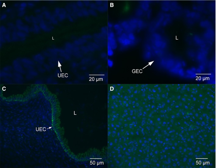
Figure 3.

Control images for immunofluorescence micrographs. (A) Negative control anti‐mouse IgG staining in uterine epithelium from early pregnant dunnart. Only blue nuclei staining present. (B) Anti‐mouse IgG staining in glandular epithelium from late pregnant dunnarts. Only blue nuclei staining present. (C) Positive control rat uterine epithelium from day 1 of pregnancy. E‐cadherin staining is localised to the cytoplasm and lateral plasma membrane of the uterine epithelium. (D) Positive control rat liver shows cytoplasmic E‐cadherin staining throughout the cells.
