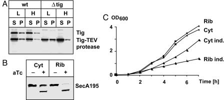
Fig. 3.
TDP with cytoplasmic and ribosome-attached SecA. (A) Cellular localization of TEV protease. Wild-type (wt) and Δtig cells expressing Tig144-TEV protease were fractionated into cytoplasmic (S) and ribosomal fractions (P) by using low (L) and high (H) salt conditions for Western blotting. Trigger factor was visualized by using anti-trigger factor antibodies and Tig144-TEV protease by anti-TEV protease antibodies. (B) Proteolysis of SecA195 by cytoplasmic and ribosomal TEV protease. Western blot analysis was performed with anti-TEV antibody. Samples were taken after 5 h of growth in NZA medium. Expression of TEV protease was induced with 100 ng/ml aTc. (C) Growth of isogenic strains expressing either cytoplasmic (Cyt) or ribosome-attached (Rib) TEV protease. Cultures were grown with or without inducer (ind.) (100 ng/ml aTc).

