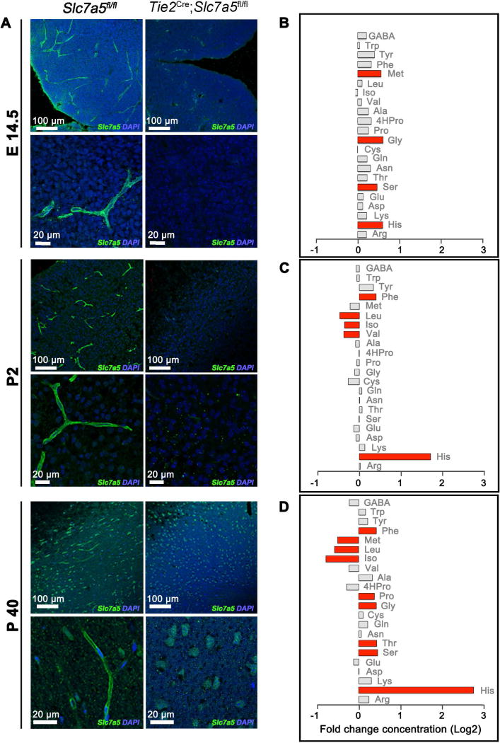
Figure 1. Slc7a5 mediates BCAA flux at the BBB.
(A) Representative images showing Slc7a5 (green) localization at the BBB in control animals (Slc7a5fl/fl, left) and its complete deletion in endothelial cells of the BBB in Cre positive mice (Tie2Cre;Slc7a5fl/fl, right). Immunostainings were performed in cortical slices at embryonic day 14.5 (E14.5 top), postnatal day 2 (P2, middle) and adulthood (>P40, bottom). Nuclei were stained with DAPI (blue). (B–D) Brain amino acid levels in Tie2Cre;Slc7a5fl/fl mice at E14.5 (B), P2-14 (C) and >P40 (D). Levels of amino acids were normalized on protein concentration and shown as fold change (log2 transformed) to levels in age-matched controls. In red are represented the amino acids with a fold change >1.3 and P value <0.05 (n>4 mice/genotype/time point).
See also Figure S1, S2 and Table S1.

