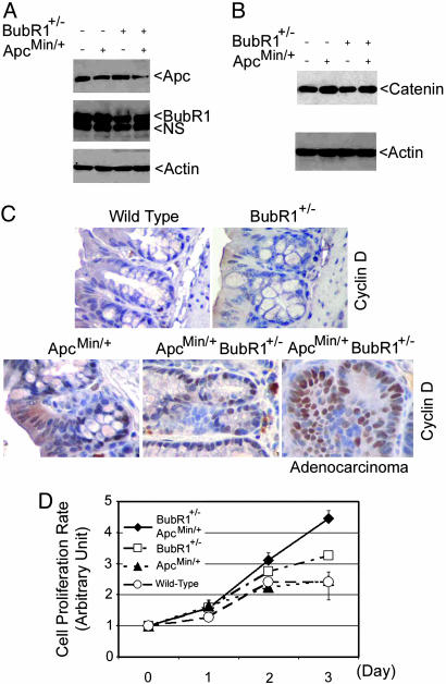
Fig. 2.
BubR1+/–ApcMin/+ MEFs proliferate at an accelerated rate. (A) Paired MEFs were lysed and an equal amount of cell lysates were blotted for Apc, BubR1, or β-actin. Arrow NS denotes a nonspecific cross-reactive band. (B) Equal amount of cell lysates from MEFs of various genotypes were blotted for β-catenin and β-actin. (C) Sections of normal colons from mice of various genotypes were subjected to immunohistochemical studies after staining with IgGs to cyclin D1. A typical adenocarcinoma section from BubR1+/–ApcMin/+ mice that was stained with cyclin D1 is also presented. (D) MEFs of various genotypes were subjected to cell proliferation assays using the 3-(4,5-dimethylthiazol-2-yl)-2,5-diphenyl tetrazolium bromide (MTT) method. The data are summarized from three independent experiments.

