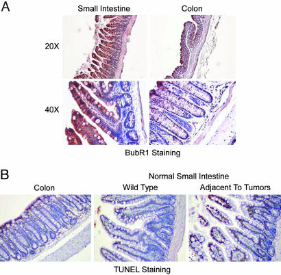
Fig. 3.
Analysis of BubR1 expression and apoptosis in small intestines by immunohistochemistry. (A) Sections of paraffin-embedded small intestine and colon from WT mice were stained with antibody to BubR1. Representative images at various magnifications are presented. Samples from at least three mice were examined. (B) Sections of paraffin-embedded small intestine and colon samples from BubR1+/–ApcMin/+ mice were examined for apoptosis by using a TUNEL kit. Representative images from three independent samples are presented.

