
FIGURE 4. Phosphorylation of myofilament cTnI and cMyBP-C residues 4 days after gene transfer.
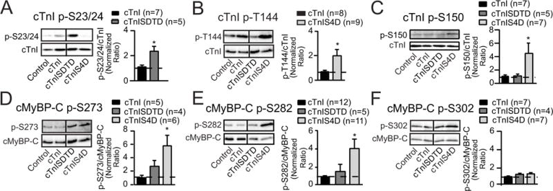
Representative immunoblots (left panels) and quantitative analysis (right panels) of phosphorylated cTnI -S23/24 (p-S23/24; A), -T144 (p-T144, B) and -S150 (p-S150, C) relative to total cTnI expression (lower panels) in controls and myocytes expressing cTnI, cTnISDTD or cTnIS4D 4 days post-gene transfer. The ratios of phosphorylated/total cTnI in each panel are normalized to control ratios, which are set to 1.0 (dotted line in each graph) for each quantitative analysis (30). The ratios in cTnISDTD- and/or cTnIS4D-expressing myocytes were compared to myocytes expressing cTnI after gene transfer, with statistical significance set at p<0.05 (*). This Western analysis also shows the expected reduction in p-S23/24 and p-T144 after replacement with cTnIS4D and cTnISDTD, respectively. Due to this reduction, p-S23/24 in cTnIS4D- and p-T144 in cTnISDTD- expressing myocytes were not quantitatively analyzed in their respective panels. Representative immunoblots shown in panels D–F show site-specific phosphorylation of cMyBP-C in the same groups of day 4 myocytes shown in A–C. Immunoblots (left panels) and quantitative analysis (right panels) are shown for phosphorylated cMyBP-C -S273 (p-S273, D), -S282 (p-S282, E), and -S302 (p-S302, F). Detection of the specific phosphorylated residue (left panels) is shown in the upper blot and total cMyBP-C in the lower panel. Quantitative analysis was performed using the normalized phosphorylated/total cMyBP-C ratio in each group, with the control ratio set to 1.0 (indicated by dashed line). Results in each panel were compared to cTnI expressing myocytes, with statistical significance set at p<0.05 (*). Vertical black lines shown in Western blots in this figure and in Figures 5 and 6 indicate a separation between cTnI and cTnISDTD on the same blot.