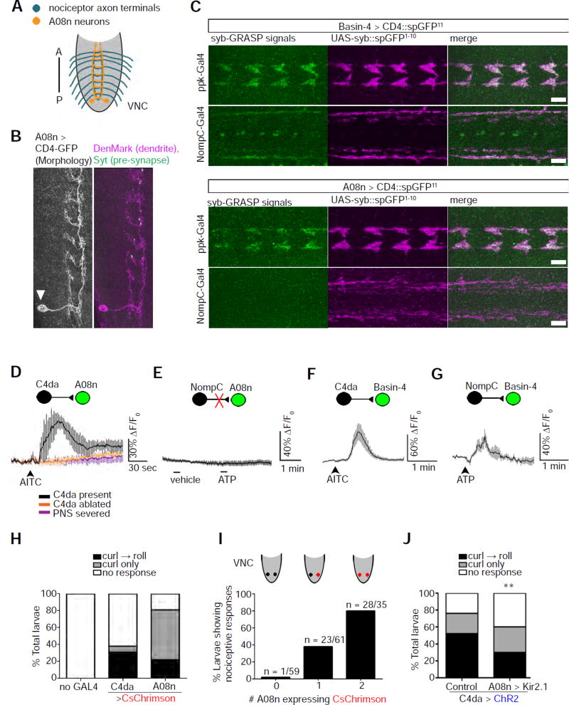
Figure 2. A08n neurons are specific postsynaptic targets of nociceptors.
(A) A schematic of the axon projections of nociceptors (blue) and the neurites of A08n within the VNC (orange). A: anterior; P: posterior.
(B) The majority of A08n neurites are dendrites. A single A08n neuron labeled by the FLP-out technique. The soma (arrowhead) of the A08n neuron is located near the posterior end of the VNC. A08n neurites in close proximity to C4da axon terminals are mostly positive for the dendrite marker, DenMark, and contain scattered spots that are positive for the axonal marker, Synaptotagmin (Syt). Scale bar: 10 µm.
(C) A08n neurons are synaptic partners of C4da, but not NompC-expressing mechanosensory, neurons. The synaptobrevin (syb)-GRASP technique was used to detect synaptic connections between neurons. SpGFP11 expressed in Basin-4 neurons produces syb-GRASP signals (green) with spGFP1–10 (magenta) expressed in either nociceptors (ppk-GAL4) or mechanosensors (NompC-GAL4). In contrast, spGFP11 expressed in A08n neurons only produces syb-GRASP signals with spGFP1–10 in nociceptors. The discrete GFP signals along the VNC midline are artifacts that show up in the absence of spGFP11 and can be observed with various antibodies. Scale bar: 10 µm.
(D) AITC activates A08n neurons specifically through C4da nociceptors, as shown by calcium imaging. A schematic is included to show pre- and post-synaptic neurons. The neuron that was recorded by GCaMP calcium imaging is shown in green. Traces show the averages of responses. No response was observed in the absence of C4da neurons (“C4da ablated”) or with the severing of nerves connecting the VNC with the PNS (“PNS severed”). n = 3, 4, and 4 in “C4da present”, “C4da ablated”, and “PNS severed”, respectively.
(E) Stimulation of NompC-expressing mechanosensors does not activate A08n neurons. Mechanosensors expressing P2X2 were stimulated by 1 mM ATP for Ca2+ imaging. n = 10 neurons (5 larvae).
(F) Stimulation of C4da nociceptors activates Basin-4 neurons. Note that AITC does not activate NompC-expressing mechanosensory neurons (data not shown). n = 9 neurons (5 larvae).
(G) Stimulating NompC-expressing mechanosensors activates Basin-4 neurons. n = 8 neurons (4 larvae).
(H) Activation of C4da or A08n neurons by CsChrimson elicits nociceptive response. n = 90 larvae for each group. Note that activation of A08n neurons mostly results in abrupt body curling.
(I) Larvae expressing CsChrimson in one or two A08n neurons exhibit an increase in nociceptive behavior. Results combine curling and rolling. Most of the responders showed curling only. Sample numbers are indicated in the graph.
(J) Larvae with silenced A08n neurons show a significant reduction in nociceptive response. Nociceptors are activated by ChR2. A08n neurons were silenced by Kir2.1 expression with the GMR82E12-Gal4 driver. The control group lacks the driver. n = 100 larvae for each group.
(See also Figure S3.)

